รายละเอียดเพิ่มเติม
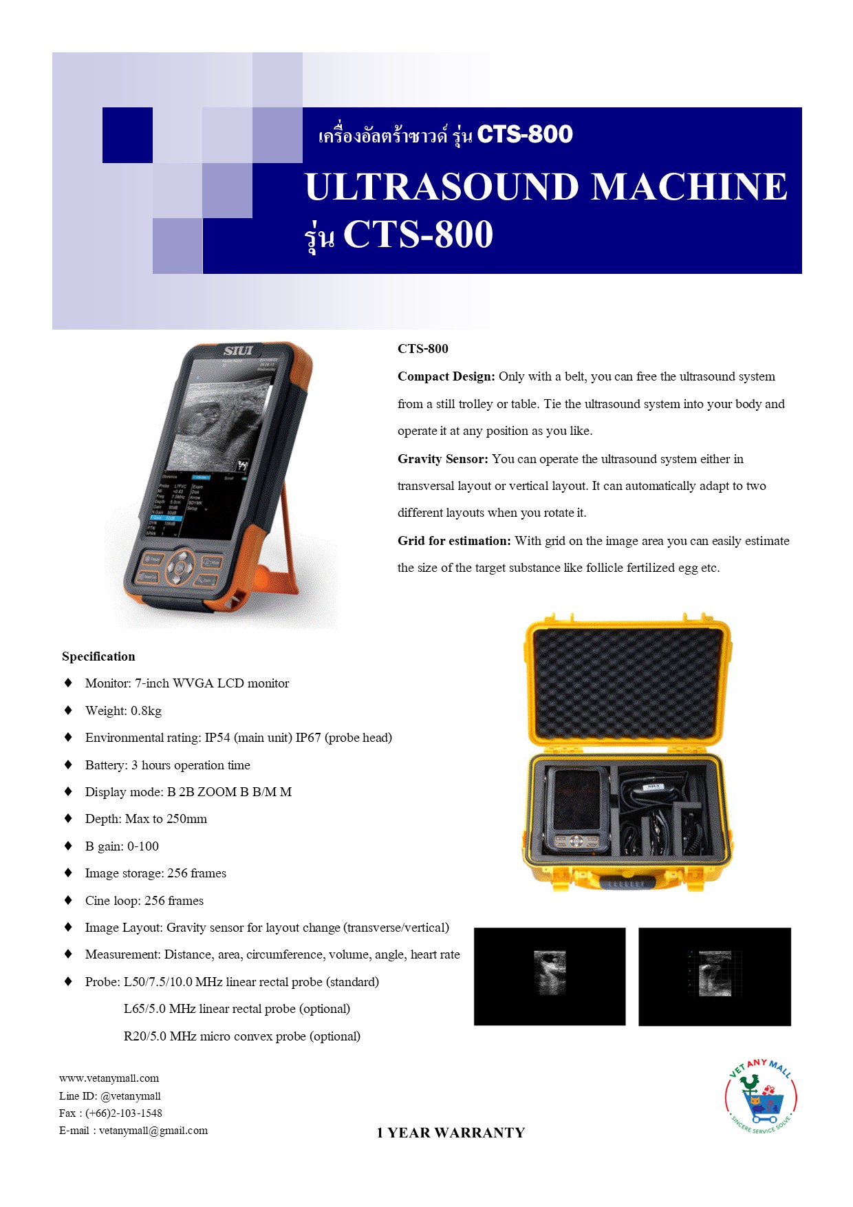
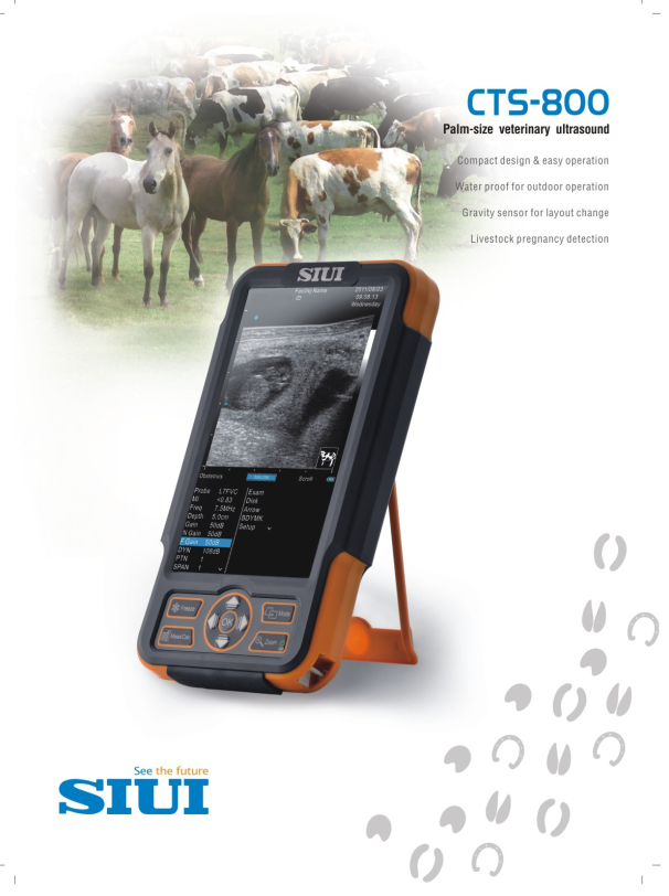
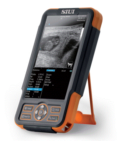
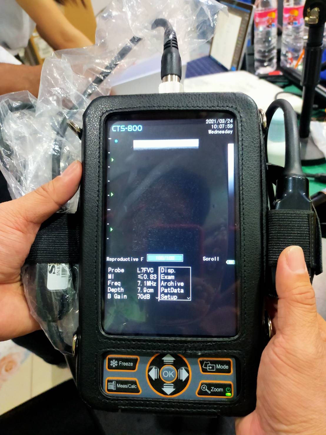
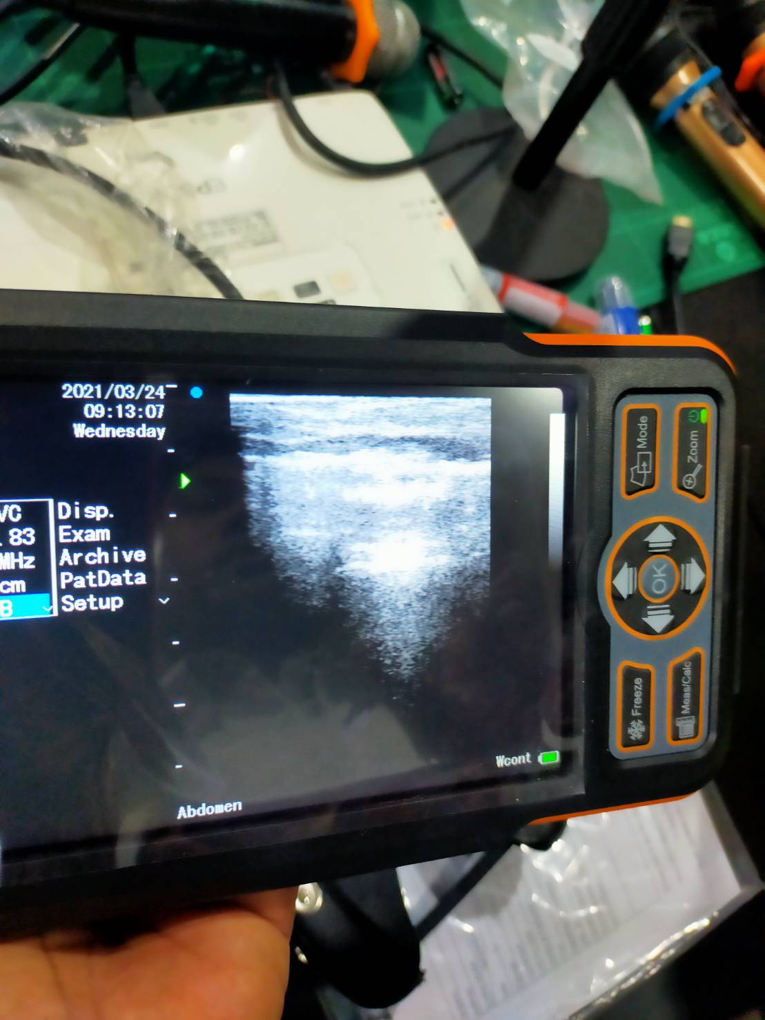
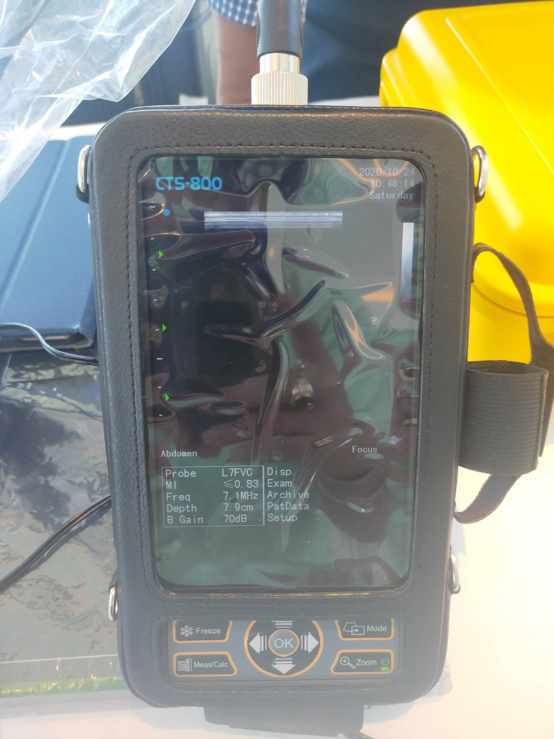
CTS-800
——Palm-size veterinary
ultrasound
Appearance
– Palm size design
– 7-inch LCD monitor
– Visual Angle: 32°
– Resolution : 800 × 480
– One active probe connectors
– Built-in lithium battery
– Working hour: over 3 hrs
– Charging time: less than 4 hrs
– Environmental rating:
IP 54 (main unit)
IP 67(probe head)
Probe
Transducer Types
– Electronic convex probe
– Electronic micro convex probe
– Electronic linear probe
– Electronic trans rectal probe
Probe Mode
– L7FVC Linear rectal probe
– L5FVC Linear rectal probe
– C3FC Convex probe
– C5FC Micro-convex probe
– L1FC Linear probe (back fat)
Technology
Applications
– Bovine
– Equine
– Ovine
– Porcine
Highlight
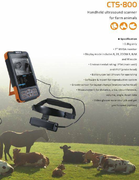
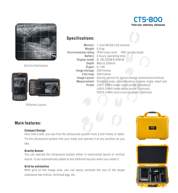
– Gravity Sensor
– Grid for estimation
– Battery
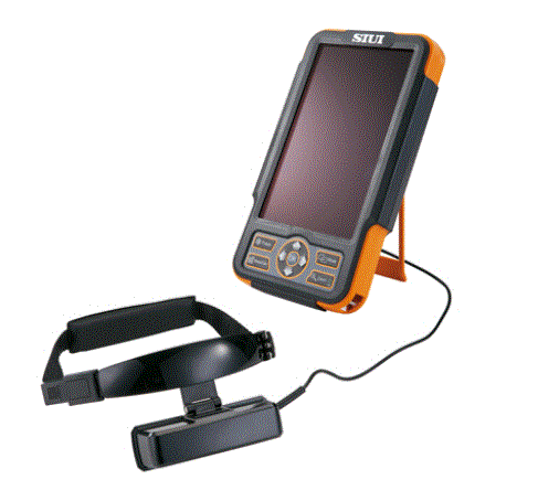
– Video glasses (Optional)
Display mode
– B mode
Product data
2 CTS-800 | www.siui.com
– 2B mode
– B/M mode
– M mode
– Zoom B mode
Zoom
– Real time zooming
– 3 Steps: ×2.0, ×3.0, ×4.0
– Selectable zooming position
– Zoom frozen
– 3 Steps: ×2.0, ×3.0, ×4.0
Focus
– Continuous dynamic focus
– Dynamic apodization
– Dynamic aperture
– 1~4 selectable transmit focus
– Acoustic lens focus
Memory
– Cine-memory
B-mode (max.256 frames)
M-mode (max.2550 seconds)
– 16 frames instant image storage
– More than 100 BMP/1000 JPG Image
storage
– Memory card 256M
Imaging Processing
2D mode
– Gain: 0~100
– Depth: 1.6~24.4cm
– Frequency: 5 steps
– Dynamic range adjustable: 36~180dB
– Edge enhancement: 0~4
– Smooth: 0~3
– Nanoview: 0~6
– Persistence: 0~7
– Chroma: 0~8
– Grayscale: 0~23
– Power: -∞~0dB, 0~100%
– Scan angle: 20°~72°
– Line density: Auto
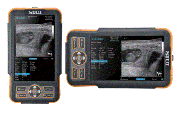
– Inversion: left/right, up/down, rotate
M mode
– Gain:0~100
– Sweep speed: 4 steps
Measurement & Calculation
Measurement
2D mode (General)
– Distance
– Trace (area)
– Ellipse (area)
– Biplane (volume)
– Ellipse (volume)
– Simpson (volume)
– Sphere (volume)
– Angle (general)
– Area ratio (trace)
– Area ratio (ellipse)
– % area reduction (trace)
– % area reduction (ellipse)
– % dima.reduction
– Histogram
– LDW volume (3 lines)
M mode
– Time
– Multiple distance
– Heart rate
– Slope
Calculation
Abdomen
Product data
3 CTS-800 | www.siui.com
– Liver
Long Left Lobe
Anteroposterior Left Lobe
Angle Left Lobe
Obli R Lobe
Anteroposterior Right Lobe
Angle Right Lobe
Portal Vein
IVC (Inferior Vena Cava)
SMA (Superior Mesenteric
Artery)
CELA (Celiac trunk)
AO (aortaventralis)
– Spleen
Length
Anteroposterior
Spleen artery
Spleen vein
– Gallbladder
Length
Anteroposterior
Transverse
Wall
CBD (Common bile duct)
LHD (Left hepatic duct)
RHD (Right hepatic duct)
– Pancreas
Head
Body
Tail
MPD(Main pancreatic duct)
– AO
% Dima.reduction
% Area reduction
Urology
– Kidney
Length Left Kidney
Anteroposterior Left Kidney
Transverse Left Kidney
Left Renal Artery
Length Right Kidney
Anteroposterior Right Kidney
Transverse Right Kidney
Right Renal Artery
– Ureter
Left
Right
– Bladder
Length
Anteroposterior
Transverse
Obstetrics
– GSD (gestation sac diameter)
– CRL (crown-rump length)
– BPD (biparietal diameter)
– HD (head diameter)
– TD (transverse trunk diameter)
– BD (Body diameter)
– Growth charts
Reproductive M
– Prostate
Volumen
PSAD (Prostate specific antigen
Density)
– Testes
Long Left Testis
Anteroposterior Left T Testis
Transverse Left T Testis
Long Left Epididymis
Anteroposterior Left Epididymis
Long Right Testis
Anteroposterior Right Testis
Transverse Right Testis
Product data
4 CTS-800 | www.siui.com
Long Right Epididymis
Anteroposterior Right Epididymis
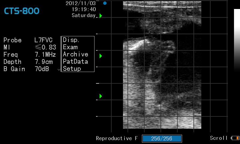
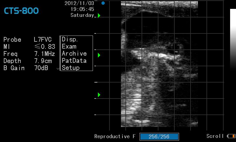
Reproductive F
– Uterus
Length
Anteroposterior
Transverse
Endometrium
– Cervix
Length
Anteroposterior
Transverse
– Ovary
Length Left
Anteroposterior Left
Transverse Left
Length Right
Anteroposterior Right
Transverse Right
– Follicle
Volume 1
Volume 2
Volume 3
Volume 4
Volume 5
Volume 6
Volume 7
Volume 8
Volume 9
Volume 10
Cardiology
– RVAWd (Right ventricular anterior
wall diastolic)
– RVd (Right ventricle diastolic period)
– RVOT (Right ventricular outflow
tract)
– AO (Aorta)
– LA (Left atrium)
– IVSd (Inter-ventricular septum in
diastolic period)
– LVd (Left ventricle in diastolic period)
– LVPWd (Diameter of left ventricle
posterior wall in diastolic period)
– RVAWs (Right ventricular anterior
wall systolic period)
– RVs (Right ventricular systolic period)
– IVSs (Inter-ventricular septum in
systolic period)
– LVPWs (Diameter of left ventricle
posterior wall in systolic period)
– RA (right ventricular)
– IVC (Inferior vena cava)
– PA (Great artery short axis view)
Physical Features
Connectivity
– Video out port
– 1 USB port
– 1 RJ-45
– AC power input port
Dimension
– Gross dimension:
– 440 mm (H) X 340 mm (W) X 185 mm
(D)
– Net dimension:
– 225 mm (H) X 128 mm (W) X 45 mm
(D)
Weight
– Gross weight : 7kg (2 probes and
carry case)
– Net weight : 0.8kg
รายละเอียดคุณลักษณะเฉพาะ
เครื่องตรวจอวัยวะภายในด้วยคลื่นเสียงความถี่สูงชนิดมือถือ
(Black White Handheld Ultrasound)
- คุณสมบัติทั่วไป
- เป็นเครื่องตรวจอวัยวะภายใน ด้วยคลื่นเสียงความถี่สูงระบบดิจิตอล แสดงภาพขาว ดำพร้อมอุปกรณ์
- ใช้กับไฟฟ้า AC 100V to 240V และมีแบตเตอร์รี่ภายในตัวเครื่อง ชนิด ลิเทียม ใช้งานเครื่องฯได้ ไม่ต่ำกว่า 3 ชั่วโมง
- คุณสมบัติเฉพาะ
- รับส่งคลื่นเสียงแบบปรับจุดโฟกัสได้ (Continuous Dynamic Focus) โดยช่องปล่อยคลื่นเสียงปรับได้ทางอิเล็กทรอนิกส์ (Dynamic Aperture) ผ่านทางจุดเลนส์รวมคลื่นเสียง (Acoustic Lens Focus)
- มีจอภาพแสดงผลชนิด LCD ขนาดไม่น้อยกว่า 7 นิ้ว มีความละเอียดไม่น้อยกว่า 800 x 480 จุด
- มีขนาดเล็ก (Palm Size) ถือด้วยมือข้างเดียวได้ และมีมุมมองภาพ 32 องศา หรือกว้างกว่า
- ใช้เวลาในการชารจ์ประจุ ไม่เกิน 4 ชั่วโมง
- ตัวเครื่อง มีความสามารถที่จะป้องกันฝุ่น และละอองน้ำที่ตกกระทบตัวอุปกรณ์ได้จากทุกทิศทาง ตามมาตราฐาน IP 54 หรือดีกว่า
- มีฟังค์ชั่นแสดงภาพได้ ดังนี้
- B Mode
- 2B Mode
- B/M mode
- M Mode
- Zoom B mode
- ขยายภาพได้ไม่น้อยกว่า 3 ระดับ ที่ 2, 3, 4 เท่า
- ปรับความลึกได้ตั้งแต่ 2 – 24 ซ.ม.
- มีอัตราขยายสัญาณในช่วงกว้าง (Dynamic Range) ตั้งแต่ 36 – 180 dB
- สามารถปรับการแสดงผลได้ทั้งแนวตั้งและแนวนอน
- คุณสมบัติการตรวจใน 2D mode
- ปรับอัตราขยายภาพได้ (GAIN) ตั้งแต่ 0 – 100 dB หรือ ดีกว่า
- ปรับความถี่ได้ 5 ความถี่
- ปรับความคมชัดขอบภาพ (Edge Enhancement) ได้ 5 ระดับ
- ปรับความนุ่มนวลของภาพ (Smooth) ได้ 4 ระดับ
- ปรับระดับกำจัดสัญญาณรบกวน (NanoView) ได้ 7 ระดับ
- คุณสมบัติการตรวจใน M Mode
- ปรับอัตราขยายภาพได้ (GAIN) ตั้งแต่ 0 – 100 dB หรือ ดีกว่า
- ปรับค่าความเร็วในการกวาดภาพ ได้ 4 ระดับ
- คุณสมบัติการวัด
- Distance
- Trace
- Ellipse
- Bi-Plane
- Ellipse
- คุณสมบัติการเก็บภาพในหน่วยความจำ
- ดูภาพ 2D ย้อนหลังได้ 256 ภาพ
- ดูภาพ M Mode ย้อนหลังได้ 2,550 วินาที
- บันทึกภาพ 2D ได้ไม่น้อยกว่า 1000 ภาพ แบบ Jpeg
- มีความจุของหน่วยความจำ ที่ 256 MB
- อุปกรณ์ประกอบมีดังนี้
- หัวตรวจ Linear Rectal 1 หัวตรวจ
- กระเป๋าหิ้วสะพายเครื่อง 1 ชุด
- กล่องใส่แบบพลาสติก 1 ชุด
- ชุดชารจ์ประจุไฟบ้าน 1 ชุด
- ชุดชารจ์ประจุในรถยนตร์ 1 ชุด
- เงื่อนไขเฉพาะ
- บริษัทต้องรับประกันเครื่องมือทุกชิ้นส่วน 2 ปี
- มีคู่มือการใช้งาน ภาษาไทย และ ภาษาอังกฤษ อย่างละ 1 ชุด
- ผลิตภัณฑ์ในประเทศเอเซีย
คลิปการใช้งาน VR glass 3D Ultrasound (Optional)
add more 25,000 bath ซื้อเพิ่ม 25,000 บาท

